
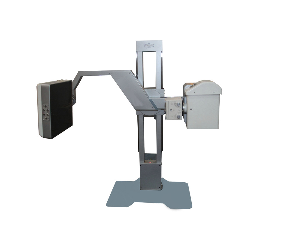
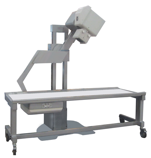
Complex X-ray diagnostic KRD «INDIascan-01»
X-ray Diagnostic Complex KRD INDIascan-01 with Cabin and Tomosynthesis Mode is designed to perform full-field digital radiography and digital tomosynthesis of the chest. The system provides high-quality visualization of the lungs, heart, and other thoracic anatomical structures, facilitating the early detection of tuberculosis, oncological diseases, cardiovascular disorders, and other pathologies of the chest organs.
The X-ray protective cabin in the complex provides reliable protection of the operator from radiation, facilitates the preparation of the room and further operation. It has a lead equivalent of not less than 1.5 mm Pb.
Increased diagnostic sensitivity of the complex is ensured by the presence of the X-ray digital tomosynthesis (DT) mode. X-ray digital tomosynthesis is a new examination method in radiology that is rapidly developing. In terms of the simplicity of performing examinations, DT is close to digital radiography (DR), and in terms of the effectiveness of the examination, it is close to computed tomography (CT). At the same time, the radiation load on patients is an order of magnitude lower than during CT examination. It does not exceed 1 mSv, which allows the use of the DT mode for preventive examinations.
DT is particularly effective in detecting early stages of tuberculosis, oncology, viral diseases, and pathologies of the chest organs.
Description
Advantages:
- Diagnostic sensitivity approaching ST.
- The radiation dose during the examination does not exceed 1 mSv.
- The device allows you to obtain clear high-contrast radiographic and tomosynthesis images.
- Robotic mechanics with microprocessor control.
- The range of vertical movement of the traverse is more than 50 cm.
- The use of a modern high-frequency power supply (100 kHz) with a power of up to 16 kW.
- A progressive high-tech solution with the organization of the production process of the examination: the X-ray technician performs remote automated control from a single integrated console, outside the irradiation zone.
The design of the X-ray Diagnostic Complex INDIascan-0 makes it possible to ensure:
- high-quality implementation of radiography and tomosynthesis;
- highly efficient and productive work during mass examinations (up to 40,000 examinations per year);
- examination of patients with non-standard build, height, disabled people, etc., reducing discomfort and eliminating the risk of injury;
- fixed emitter-receiver distance of 1400 mm (minimizing projection distortions).
- convenient and universal software with a tomographic interface.
Specifications
|
|
|
|
|
Radiography Mode |
Supported |
|
|
1 |
Generator power |
16 kW |
|
2 |
Tube voltage (kVp) range |
50–120 kV |
|
3 |
Tube current range |
10–100 mA |
|
4 |
Digital detector active area |
43 × 43 cm |
|
5 |
Spatial resolution |
3.7 lp/mm |
|
6 |
Dynamic range |
16-bit |
|
7 |
Source-to-image distance (SID) |
1400 mm |
|
Digital Tomosynthesis Mode |
Supported |
|
|
8 |
Scan type |
Horizontal |
|
9 |
X-ray tube travel |
700 mm |
|
10 |
Detector travel |
200 mm |
|
11 |
Number of projection images |
65–120 |
|
12 |
Scan time |
5–10 s |
|
13 |
Number of reconstructed slices |
100–250 |
|
14 |
Spatial resolution (Tomosynthesis mode) |
2.0 lp/mm |
|
15 |
Dynamic range |
16-bit |
|
16 |
Generator power |
5 kW |
|
17 |
Tube voltage (kVp) range |
50–100 kV |
|
18 |
Tube current range |
10–40 mA |
|
Operator/Radiologist Workstation |
Included |
|
|
19 |
Image communication standard |
DICOM 3.0 |
|
20 |
Workstation type |
Personal Computer (PC) |
|
Radiation-Shielded Cabin |
Included |
|
|
22 |
Lead equivalence |
≥ 1.5 mm Pb |
|
23 |
Dimensions (W × D × H) |
1190 × 2130 × 2000 mm |
|
X-ray Tube |
Included |
|
|
24 |
Focal spot sizes |
1.0 / 2.0 mm |